Oral administration of the β-glucan produced by Aureobasidium pullulans ameliorates development of atherosclerosis in apolipoprotein E deficient mice

Aureobasidium pullulans-derived β-glucan (AP-PG) is known to be an immunomodulator.

Apolipoprotein E deficient (ApoE) mice spontaneously develop atherosclerosis.

AP-PG is effective in ameliorating development of atherosclerosis in the model mice.

AP-PG administration reduces oxidized low-density lipoprotein cholesterol levels.

Oral administration of AP-PG ameliorates vascular accumulation of macrophages.

Abstract

The Aureobasidium pullulans-produced β-glucan (AP-PG) is an immune stimulator, and believed to exhibit beneficial effects on health through its immune stimulating activity. Here, the effect of oral administration of AP-PG on high-fat diet (HFD)-induced atherosclerosis was evaluated using apolipoprotein E deficient mice, a widely used mouse model for atherosclerosis. The results demonstrated that HFD-induced development of atherosclerosis was significantly reduced in the AP-PG-treated mice when compared with that of the control mice. In serological analysis, blood levels of oxidized low-density lipoprotein cholesterol, a well-known risk factor for the development of atherosclerosis, were significantly reduced in the AP-PG-treated group of mice. Further, immunohistochemical analysis using MOMA-2 antibody showed that oral administration of AP-PG is effective in ameliorating vascular accumulation of macrophages. These data suggest the possibility that oral administration of AP-PG is effective in ameliorating HFD-induced development of atherosclerosis.

Abbreviations

ApoE

apolipoprotein E

AP-PG

A. pullulans-produced β-glucan

CAD

coronary artery disease

HDL

high-density lipoprotein

HFD

high-fat diet

LDL

low-density lipoprotein

ox-LDL

oxidized low-density lipoprotein

Keywords

β-glucan
Aureobasidium pullulans
Atherosclerosis

1. Introduction

Black yeast, Aureobasidium pullulans, extracellularly produces a soluble β-glucan (AP-PG) consisting of a β-(1,3)-linked main chain and β-(1,6)-branched glucose residues under certain conditions (Moriya et al., 2013), and the β-glucan-containing A. pullulans-cultured fluid is consumed as a supplemental food promoting health. The A. pullulans-produced β-glucan is highly branched with β-(1,6)-linked glucose residues, and is known to be structurally distinct from other organism derived β-glucans. It is believed that the A. pullulans-produced β-glucan is effective in promoting health, and has anti-tumour (Kataoka-Shirasugi et al, 1994Kimura et al, 2006), anti-allergy (Kimura, Sumiyoshi, Suzuki, Suzuki, & Sakanaka, 2007), and anti-infectious disease (Muramatsu et al, 2012Muramatsu et al, 2014Yatawara et al, 2009) effects.
In this study, we demonstrated that oral administration of AP-PG is effective in ameliorating HFD-induced development of atherosclerotic lesions in apolipoprotein E (ApoE) deficient mice which spontaneously develop atherosclerotic lesions (Plump et al, 1992Zhang et al, 1992). In the serological analysis, the blood level of oxidized LDL cholesterol increased after feeding with HFD, and this increment was significantly decreased in AP-PG administered mice compared with that in the control mice. These results suggest that oral administration of AP-PG is effective in preventing HFD-induced development of atherosclerosis.

2. Materials and methods

2.1. Mice

Specific pathogen-free 8 week old ApoE deficient male mice of C57BL/6N background were purchased from Jackson Laboratory (Bar Harbor, ME, USA). All animal experiments were performed in accordance with the guidelines of the Bioscience Committee of Hokkaido University and were approved by the Animal Care and Use Committee of Hokkaido University (permit number 13–0179).

2.2. Preparation of A. pullulans-derived β-glucan

For preparation of the A. pullulans-derived β-glucan (AP-PG), A. pullulans-cultured fluid was subjected to diatomaceous earth filtration to remove cell debris, and this was followed by removal of low molecular weight components by ultrafiltration with a cut-off molecular weight of 20,000 Da (Q2000; Advantec, Tokyo, Japan). Subsequently, β-glucan was precipitated with ethanol at a concentration of 80% and used in this study. The purity of the prepared β-glucan was estimated to be above 99% using high performance liquid chromatography (HPLC). The low molecular weight fraction enriched by ultrafiltration was used as the control in this study. The removal of β-glucan from the control fraction was confirmed using HPLC, and the concentration of β-glucan was estimated to be below 1%.

2.3. Measurement of cholesterols and triacylglycerol

Serum concentrations of cholesterol and triacylglycerol were measured using commercially available kits (Triglyceride E-test Kit and Cholesterol E-test Kit; Wako Pure Chemical, Osaka, Japan). Blood levels of high-density lipoprotein (HDL) cholesterol and oxidized low-density lipoprotein (ox-LDL) were monitored using the HDL cholesterol E-test Wako (Wako) and an ELISA Kit for Oxidized Low Density Lipoprotein (USCN Life Science Inc, Wuhan, China), respectively. Each procedure was performed according to the manufacturer’s protocols, and the blood low-density lipoprotein cholesterol levels were calculated by the following formula: LDL cholesterol = total cholesterol − HDL cholesterol − (triacylglycerol × 0.2).

2.4. Histochemical and immunohistochemical analyses

The MOMA-2 antibody was purchased from Serotec, Oxford, UK, and Sigma-Aldrich, St. Louis, MO, USA. The procedure for the immunohistochemical staining, haematoxylin and eosin (HE) staining, Oil-O-Red staining is described elsewhere (Matsui et al., 2003).

2.5. Image analysis and statistical analysis

The analysis of the histochemical images was performed using Adobe Photoshop (Adobe Systems, San Jose, CA, USA). To check for significant differences between the indicated pairs of data, the two-tailed unpaired Student’s t-test was used in this study.

3. Results

3.1. Oral administration of AP-PG is effective to ameliorate development of atherosclerosis in ApoE deficient mice

To evaluate the effects of orally administered AP-PG to HFD-induced arteriosclerosis, we used ApoE deficient mice. An outline of the experimental design is shown in Fig. 1. The AP-PG in the drinking water was diluted to a concentration of 200 µg/ml, and the AP-PG dose under this condition was estimated to be about 2 mg/kg as evaluated by the drinking water intake. After one week of pretreatment with AP-PG, the mice were given the HFD for 16 weeks, and the administration of AP-PG was continued till the end of the experiment. The body weight and food intake changes of the mice are shown in Supplemental Fig. S1. The results show that the oral administration of AP-PG did not significantly affect the food intake and body weight of the mice.
  1. Download: Download full-size image

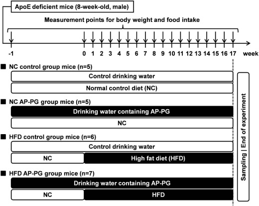
Fig. 1. Experimental design of this study.

Next, the effect of orally administered AP-PG on the HFD-induced development of atherosclerosis was assessed by Oil-Red-O staining. The image analysis and microphotographs of Oil-Red-O-stained aortae are shown in Fig. 2A and B, respectively. These results show that Oil-Red-O stained aortic atherosclerosis was significantly augmented by the HFD in the control group mice; the HFD-induced aortic atherosclerosis was significantly reduced in the AP-PG administered group of mice. We also evaluated the development of atherosclerotic lesions around the aortic roots. The HFD induced-atherosclerosis around the aortic roots was significantly lower in the AP-PG administered mice than in the control mice (Fig. 2C and D). These results indicate that the oral administration of AP-PG is effective to ameliorate HFD-induced development of atherosclerosis in ApoE deficient mice.
  1. Download: Download full-size image

Fig. 2Oral administration of AP-PG effectively prevented high-fat diet-induced development of atherosclerotic plaque in ApoE deficient mice. (A and B) At the end of the period of the experiment, the main artery was isolated from the mice and atherosclerotic plaque formation was evaluated using Oil-O-Red staining. Data represent the image analysis results (A) and histopathological specimen images (B). Scale bars indicate 1 cm. (C) Image analysis of the atherosclerotic lesions on the aortic root sections. (D) Microphotographs of the histology of the aortic root sections stained with Oil-O-Red. Scale bars indicate 50 µm. Error bars indicate the standard deviation. NC: normal control diet. HFD: high-fat diet. n.s.: not significant.

3.2. Oral administration of AP-PG reduces blood levels of oxidized low-density lipoprotein cholesterol

To evaluate effects of orally administered AP-PG on the lipid metabolism, the blood levels of cholesterol and triacylglycerol were measured using commercially available kits. The results show that blood cholesterol and triacylglycerol levels were not significantly different for the HFD AP-PG and HFD control groups (Fig. 3A and B, respectively). Next, the blood levels of high-density lipoprotein (HDL) cholesterol and low-density lipoprotein (LDL) cholesterol were quantified. The results show that the amounts of blood HDL and LDL cholesterols were also not significantly changed by the oral administration of AP-PG (Fig. 3C and D, respectively). Finally, the blood level of oxidized LDL (ox-LDL) cholesterol was measured. The results show that the blood ox-LDL cholesterol level was significantly reduced in the HFD AP-PG group mice, compared with that in the HFD control group (Fig. 3E).
  1. Download: Download full-size image

Fig. 3. Evaluation of the effects of orally administered AP-PG on blood cholesterol levels (A–E). At the end of the period of the experiment, plasma levels of total cholesterol (A), triacylglycerol (B), high-density lipoprotein (HDL) cholesterol (C), low-density lipoprotein (LDL) cholesterol (D), and oxidized low-density lipoprotein (ox-LDL) cholesterol (E) in the mice were measured as described in the Materials and Methods section. Error bars indicate the standard deviation. NC: normal control diet. HFD: high-fat diet. n.s.: not significant.

3.3. Macrophage accumulation in vascular walls is reduced by oral administration of AP-PG

To evaluate the effects of orally administered AP-PG on macrophage accumulation and foam cell formation, sections of aortic roots were immunohistochemically analysed using MOMA-2 antibody. The image analysis results and histological microphotographs are shown in Fig. 4A and B, respectively. These results show that the macrophage accumulation as defined by the MOMA-2 positive areas was significantly smaller in the HFD AP-PG group mice than in the HFD control group mice (Fig. 4A and B). The data suggest that oral administration of AP-PG is effective to ameliorate vascular accumulation of macrophages and foam cell formation from the macrophages.

4. Discussion

This study demonstrates that oral administration of AP-PG is effective to ameliorate the development of atherosclerosis caused by HFD in ApoE deficient mice. Serological analysis indicated that the blood ox-LDL level was significantly lower in the AP-PG group mice than in the control group mice (Fig. 3E). The blood ox-LDL level is closely related to the development of atherosclerosis (Itabe, Ueda, 2007Mitra et al, 2011), and the reduction of blood ox-LDL level after oral administration of AP-PG could be hypothesized to be an important mechanism for the amelioration of HFD-induced arteriosclerosis.
In this study, to evaluate the effects of the β-glucan in A. pullulans-cultured fluid clearly, purified β-glucan (AP-PG) was prepared, and a β-glucan-free fraction of A. pullans-cultured fluid was used as the control. Although this fraction was assumed to be containing several low molecular weight components, the body weight changes of the control group mice were almost the same as that of the mice received with water without additive (data not shown). Therefore, administration of the β-glucan-free fraction is not thought to significantly affect the results in this study.
β-glucans including AP-PG are known to be a dietary fibre, and are not absorbed in human and mouse bodies. On the other hand, the results of our preliminary experiments using radiolabelled cholesterol indicate that oral administration of AP-PG does not significantly inhibit cholesterol absorption in the small intestine under the experimental condition of this study (Aoki et al., 2015). Therefore, the influence of AP-PG as a dietary fibre on the results shown in this study is assumed to be weak, and other effects of AP-PG are thought to be important in ameliorating HFD-induced development of atherosclerosis.
Inflammation followed by oxidative stress caused by reactive oxygen species (ROS) production is closely related to ox-LDL production. Here it should be noted that there are reports of anti-inflammatory effects of β-glucan produced by A. pullulans (Kimura et al, 2007Tanaka et al, 2011). In addition, several reports have demonstrated the antioxidative effects of β-glucans derived from other organisms (Mirjanaa et al, 2013Suchecka et al, 2015Uskoković et al, 2013). Therefore, it is tempting to speculate that AP-PG has antioxidative effects, and that the anti-inflammatory and antioxidative effects of AP-PG may be involved in the reduction of blood ox-LDL levels.
The results of the immunohistochemical analysis indicated that MOMA-2 positive areas were reduced by the oral administration of AP-PG (Fig. 4), suggesting that vascular accumulation of macrophages and subsequent foam cell formation were reduced by oral administration of AP-PG. Since vascular accumulation of macrophages is closely related to the blood ox-LDL cholesterol levels (Maiolino et al, 2013Yu et al, 2013), the reduction of macrophage numbers in the vascular walls may be due to the reduction of blood ox-LDL cholesterol levels by the oral administration of AP-PG. A previous report demonstrated that orally adiministered β-glucan was able to activate lymphocytes localizing Peyer’s patch (Hashimoto, Suzuki, & Yadomae, 1991). Thus, the immune modulating activity of AP-PG is thought to be expressed by an indirect manner through the activation of small intestinal immunity. In addition, there is a report of the modulating effects of the oral administration of Sclerotinia sclerotiorum-derived β-glucan on alveolar macrophage functions (Sakurai et al., 1992), suggesting the possibility that orally administered AP-PG modulates the immune response of circulating macrophages, a source of vascular wall macrophages.
  1. Download: Download full-size image

Fig. 4Oral administration of AP-PG is effective to prevent vascular accumulation of macrophages. (A and B) Sections of aortic roots sampled from the mice were immunostained with MOMA-2. (A) Percentages of MOMA-2 positive areas were calculated by image analysis. Error bars indicate the standard deviation. NC: normal control diet. HFD: high-fat diet. n.s.: not significant. (B) Microphotographs of immunohistology sections. Scale bars indicate 300 µm.

5. Conclusions

In the present study, we demonstrated that orally administered AP-PG induced amelioration of HFD-induced atherosclerosis in a mouse model. Also, A. pullulans-cultured fluid containing β-glucan as a main component is commonly consumed as a supplemental food for long periods, and the safety as a food additive is well established. In addition, there were no negative effects on the AP-PG administered mice in this study, supporting the safety of AP-PG. However, further study is needed to establish whether AP-PG as a supplement has beneficial effects on the development of atherosclerosis in humans.