Keywords
Introduction
The cell walls of fungi evoke a powerful immunostimulatory response and have been proposed for use as potential anti-infective and antitumor drugs (Hong et al., 2003, 2004; Reynolds et al., 1980; Tzianabos et al., 1998). Both pathogenic (Candida albicans) and nonpathogenic (Saccharomyces cerevisiae) fungi share similarities in their cell wall, which is composed of an inner layer of β-glucan (60%) covalently linked to a variety of cell surface mannoproteins (40%) (Klis et al., 2001, 2002; Magnelli et al., 2002). The β-glucan is a polymer of glucose, which is mostly in the β-1,3 linkage but also contains glucose in the β-1,6 linkage. The β-1,6 glucan is estimated to be between 9%–20% of the total β-glucan (Kapteyn et al., 1999; Magnelli et al., 2002). β-1,6-glucan has an important role connecting GPI-anchored proteins to the β-1,3-glucan network (Klis et al., 2001, 2002).
Fungal β-glucan is key to immune recognition in macrophages; it stimulates phagocytosis and production of inflammatory cytokines (Brown, 2006; Brown and Gordon, 2001). This nonopsonic recognition of β-glucan is mediated mainly via Dectin-1 with cooperation of TLRs, including TLR2 (Brown, 2006; McGreal et al., 2005). Dectin-1 activity in macrophages is inhibited by both β-1,3-glucans and β-1,6-glucans, but the β-1,3-glucan, laminarin, has a strong effect and the β-1,6-glucan, pustulan, has the weakest (Brown and Gordon, 2001). Oligosaccharide microarray results also show that macrophage Dectin-1 binds specifically to β-1,3-glucans (Palma et al., 2006).
Recent reports have shown that surface mannoproteins can mask the underlying β-glucan from the receptors (Gantner et al., 2005; Wheeler and Fink, 2006). Strains of Saccharomyces cerevisiae and Candida albicans have low reactivity with either Dectin-1 or an antibody to β-glucan unless the mannoprotein layer is disrupted either by heat treatment or mutation (Wheeler and Fink, 2006). There appears to be a conserved genetic network of interacting proteins responsible for the integrity of the cell wall. Once this architecture is perturbed so that the β-glucan is exposed, the fungi interact strongly with Dectin-1 and elicit an enhanced proinflammatory response in primary mouse macrophages (Wheeler and Fink, 2006).
Early studies implicated fungal cell wall β-glucan in the stimulation of neutrophils through complement receptor 3 (CR3). CR3 has a lectin domain (Ross et al., 1985, 1987) that binds intact yeast and yeast β-glucan. Ligation of β-glucans to neutrophil CR3 increases their chemotactic capacity toward the complement fragment C5a (Tsikitis et al., 2004) and in vivo enhances neutrophil migration into the site of injury (LeBlanc et al., 2006). Although these studies provided convincing evidence for the stimulation of neutrophils by β-glucan, the exact chemical composition of the immunostimulant was unclear because the β-glucan used was a mixture of polymers. Yeast cell walls, the source of glucan in these studies, are 80% β-1,3-glucan (Magnelli et al., 2002), which led to the assumption that the β-1,3 polymer was responsible for the immunostimulatory effects. However, these cell wall preparations also contain β-1,6-glucan.
Other sources of glucan mixtures have also been shown to bind and prime neutrophil CR3, including β-glucan from barley (70% β-1,4-glucan, and 30% β-1,3-glucan) and laminarin (β-1,3-glucan with occasional β-1,6 branch points) (Thornton et al., 1996; Vetvicka et al., 1996; Xia et al., 1999). In none of these cases was a glucan of homogeneous composition compared with another for relative efficacy in stimulating immune cells for both phagocytosis and reactive oxygen species (ROS) production.
Here we show that β-1,6-glucan, the minor component of the fungal cell wall, causes a much greater stimulation of human neutrophils than β-1,3-glucan under opsonic conditions. β-1,6-glucan is more effective than β-1,3-glucan in eliciting phagocytosis, production of ROS, and elicitation of heat shock proteins by human neutrophils purified from whole blood. Neutrophil recognition of fungi is likely to be mediated by this specific sugar interaction as beads coated with β-1,6-glucan bind the complement factors C3b and its proteolytic fragment C3d better than those coated with β-1,3-glucan. This recognition is inhibited by anti-CR3 blocking antibodies, suggesting that recognition of C3b proteolytic products by CR3 mediate this enhanced phagocytosis and ROS production.
Results
Expression of Heat Shock Proteins in Neutrophils Is Induced by Exposed β-Glucan
To determine the opsonic response of neutrophils to the yeast form of Candida albicans, we performed microarray experiments using whole genome Affymetrix arrays on RNA extracted from neutrophils that had ingested the fungus. Among the genes induced in neutrophils was a family of heat shock proteins (HSPs) (The 10 kDa HSPE1, 40 kDa DNAJB1 and DNAJB9, 70 kDa HSPA1A and HSPA9B, 90 kDa HSPCA and HSPCB, and 105/110 kDa HSPH1) (see Table S1 available in the Supplemental Data available with this article online). These inductions were corroborated by quantitative real-time PCR (Figure 1A).

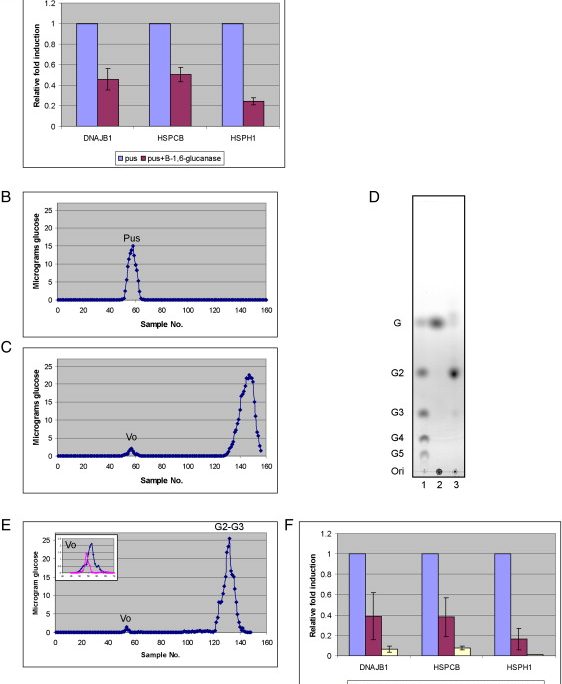
Figure 1 β-1,6-Glucan Stimulates Expression of Heat Shock Proteins in Neutrophils
The induction of HSPs was greater if Candida was first heated to unmask the underlying β-glucan (Figure 1B), suggesting that expression of HSPs is induced by the exposed β-glucan.
β-1,6-Glucan Elicits Expression of HSPs in Neutrophils
To assay which of the glucan components was responsible for induction of HSPs, we presented neutrophils with various glucan polymers. As unprimed neutrophils respond best to insoluble material (Fossati et al., 2002), we conjugated several sources of β-glucan to 6 μm polystyrene beads, which are similar in size to Candida albicans yeast form cells (5 μm). We utilized β-1,3-glucan and β-1,6-glucan purified from Candida cell walls, as well as nonfungal sources of β-1,3-glucan (laminarin) and β-1,6-glucan (pustulan), both used as standards for these polymers in previous studies (Brown and Gordon, 2001; Palma et al., 2006). The extent of coating was quantified as described in Experimental Procedures.
Beads coated with Candida-derived β-1,6-glucan were more efficient than beads coated with Candida-derived β-1,3-glucan at elicitation of HSPs in neutrophils (Figure 1C). Similar results were observed with the standards: pustulan (β-1,6-glucan) being more powerful stimulant than laminarin (β-1,3-glucan). Beads coated with pustulan elicited high levels of HSPs in neutrophils (Figure 1D), whereas beads coated with laminarin elicited only low levels of HSPs expression (Figure 1D) despite efficient coating of the beads. Glucans in the soluble form elicited only low levels of HSPs in neutrophils (Figure 1E), suggesting that neutrophils respond when the polysaccharides are presented on particles. β-glucan from barley, which is composed of β-1,3-glucan (30%) and β-1,4-glucan (70%), also did not elicit HSPs expression (Figure 1E). Beads coated with α-1,6-glucan (dextran) did not elicit any response (Figure 1E), suggesting that the neutrophil response is specific to the β configuration. Elicitation of HSPs required heat-labile serum components, as pustulan-coated beads did not elicit HSPs when opsonized with heat-inactivated (HI) serum (Figure 1D). We conclude that neutrophils express HSPs following ingestion of opsonized beads coated with β-1,6-glucan.
The Induction of HSPs by Pustulan Is Due to β-1,6-Glucan
Digestion of pustulan with an endoglucanase specific for the β-1,6-glucan linkage (Lora et al., 1995) confirmed that it consisted of β-1,6-glucan. Pustulan digested with the enzyme resulted in as much as 80% reduction in elicitation of HSPs (Figure 2A). Analysis of the digestion products of pustulan by column chromatography (see Experimental Procedures) revealed the expected peak (Figure 2 compare B to C), composed of di-and trisaccharides, as determined by TLC (G2-G3, compare lanes 2 and 3, Figure 2D). These short polymers in the G2-G3 fraction did not coat the beads efficiently (see Experimental Procedures).

Figure 2 Elicitation of Heat Shock Proteins by Pustulan Is Due to β-1,6-Glucan
In addition to the expected digestion products there was a small amount of polysaccharide (∼5% of the starting material) that was resistant to the enzyme (Figure 2C, designated as Vo) and elicited expression of the HSPs in the neutrophils. Since pustulan is reported to have a small percentage of O-acetylated β-1,6-glucan (Nishikawa et al., 1970), the small peak resistant to enzymatic digestion could be the acetylated polymer. Consistent with this interpretation, deacetylation of pustulan prior to digestion and fractionation (see Experimental Procedures) virtually abolished this minor component (Figure 2E, insert) and rendered the digested material unable to elicit HSPs (Figure 2F). The residual activity of pustulan digested with the endo-β-1,6-glucanase is therefore attributable to O-acetylated β-1,6-glucan that was resistant to the enzyme and coated the beads efficiently. Chemically acetylated laminarin (β-1,3-glucan) did not elicit any expression of HSPs (Figure S1), suggesting that the stimulation of neutrophils by β-1,6-glucan is not due to acetylation per se. Based on these results we conclude that the activity of pustulan is due to the β-1,6-glucan.
β-1,6-Glucan Mediates Efficient Phagocytosis and Production of Reactive Oxygen Species by Neutrophils
Time-lapse microscopy of neutrophils ingesting beads revealed that beads coated with pustulan (β-1,6-glucan) were more efficiently internalized than beads coated with laminarin (β-1,3-glucan) (Figure 3A, compare Ad to Ab, and compare Movie S2 to Movie S1). Neutrophils readily ingested beads when coated with pustulan. In contrast, most neutrophils ignored the laminarin-coated beads, and only a few of them ingested beads (Figure 3Ab and Movie S1, a neutrophil ingesting one bead on the upper left corner, and one neutrophil ingesting two beads on the upper right corner).

Figure 3 β-1,6-Glucan Stimulates Phagocytosis and Production of Reactive Oxygen Species in Neutrophils
Quantification of a population of neutrophils ingesting beads by Fluorescence Activated Cell Sorting (FACS) support the idea that pustulan mediates more efficient internalization of beads than laminarin, β-1,3-glucan from Candida, or β-glucan from barley, although laminarin and β-1,3-glucan from Candida promoted internalization somewhat better than uncoated beads (Figure 3B, compare panel Be to panels Bb–Bd, respectively).
Since killing of pathogens by neutrophils depends on a burst of ROS (Babior et al., 1973), we tested whether ROS were induced by either of the two glucans. ROS could not be detected in neutrophils alone or in neutrophils presented with beads that had not been treated (Figure 3C, red and green, respectively). Low levels were detected with beads coated with laminarin, β-glucan from Candida, or β-1,3-glucan from barley (Figure 3C, blue, brown, and purple, respectively). However, beads coated with pustulan stimulated the generation of significant amounts of ROS (Figure 3C, light blue). No ROS was detected by adding soluble pustulan (Figure 3C, pink). These data show that β-1,6-glucan evokes a massive production of ROS, whereas the response to β-1,3-glucan is much lower by comparison.
Complement Factor C3 Proteolytic Fragments Are Efficiently Deposited on β-1,6-Glucan
As serum is required for phagocytosis and the induction of HSPs by beads coated with β-1,6-glucan (Figure 1D), we determined whether there was a serum component that was differentially deposited on beads coated with either β-1,6-glucan or β-1,3-glucan. Beads were coated with laminarin or pustulan and opsonized as described in Experimental Procedures. Bound proteins were removed from beads and separated by SDS gel electrophoresis (see Experimental Procedures). Although a number of serum proteins bound both β-1,3-glucan or β-1,6-glucan, there were two prominent proteins that adhered more avidly to β-1,6-glucan (Figure 4A). These proteins were extracted from the gel and subjected to analysis by mass spectrometry. The peptides from these bands gave masses that identified both proteins as C3, suggesting that proteolytic fragments of C3 are deposited more avidly on β-1,6-glucan than β-1,3-glucan.

Figure 4 C3 Proteolytic Fragments Are Deposited on β-1,6-Glucan
Western analysis revealed that, indeed, β-1,6-glucan was deposited with more C3 (Figure 4B). Antibodies specific for the alpha chain detected high molecular weight bands, including bands the size of the complete C3 or C3b (105 and 115 kDa, respectively), as well as a lower molecular doublet the size of C3d (31 and 33 kDa, Figure 4Ba). Beads coated with laminarin had low levels of these C3 fragments (Figure 4Ba). Antibodies specific for the C3 beta chain revealed only the high molecular weight C3/C3b (Figure 4Bb). The 75 kDa chain of C3b or iC3b were not detected (Figure 4Bb).
To assess further the differences in C3 deposition on β-1,6-glucan as compared with β-1,3-glucan, serum was preincubated with soluble pustulan or laminarin before using it to opsonize beads coated with pustulan. Preincubation of the serum with soluble pustulan eliminated phagocytosis (Figure 5A, compare Ac and Aa) and ROS production (Figure 5B, compare red and blue) by neutrophils, suggesting that serum C3 was titrated out by the soluble β-1,6-glucan. Serum that was preincubated with an equivalent amount of soluble laminarin still mediated phagocytosis (Figure 5A, compare Ab and Aa) and ROS production (Figure 5B, compare red and green), suggesting that β-1,3-glucan did not efficiently block the interaction of C3 with beads coated with pustulan.

Figure 5 Preincubation of Serum with Soluble β-1,6-Glucan Abolishes Stimulation of Neutrophils, Whereas Soluble β-1,3-Glucan Does Not
Western analysis revealed that preincubation with soluble pustulan eliminated most of the C3/C3b and C3d deposition on the pustulan-coated beads (Figure 5C, compare 1 to 3). Preincubation with laminarin eliminated the low molecular weight C3d doublet, but retained the high molecular weight C3/C3b (Figure 5C, compare 2 to 3), suggesting that the remaining C3/C3b was mediating phagocytosis and induction of ROS (Figures 5A and 5B). Preincubation of serum with soluble pustulan but not laminarin reduced the killing of Candida albicans by 6-fold (Figure 5D). This inhibition by soluble β-1,6-glucan is consistent with the idea that complement deposition on β-1,6-glucan is required for efficient killing of the fungus.
CR3 Recognizes C3 Fragments that Are Deposited on Particulate β-1,6-Glucan
Because complement receptor 3 (CR3) is known to mediate phagocytosis of opsonized yeast and the yeast cell wall preparation zymosan, as well as ROS production (Cain et al., 1987; Ross et al., 1985), we surmised that CR3 could mediate phagocytosis of beads coated with β-1,6-glucan. Anti-human CR3 blocking antibodies reduced the extent of phagocytosis (Figure 6A, compare Ab to Aa) and ROS production (Figure 6B, compare red to green), suggesting that CR3 recognized C3b proteolytic fragments (C3d) that are deposited on particulate β-1,6-glucan.

Figure 6 CR3 Mediates β-1,6-Glucan Stimulation of Neutrophils
β-1,6-Glucan Recognition Enhances Phagocytosis of Candida albicans
β-1,6-glucan also plays an important role in the neutrophil’s recognition of whole fungal cells, which contain other polysaccharides such as chitin and β-1,3-glucan as well as many cell surface proteins. Candida albicans cells were heat killed to expose their masked β-glucan, and one sample was digested with an endo β-1,6-glucanase, and the other was incubated without the enzyme. Both samples were opsonized (see Experimental Procedures) and tested for their reactivity. The cells exposed to the enzyme (as compared with those that were not) exhibited an ∼50% reduction in phagocytosis (Figure 7A) and ROS production (Figure 7B), as well as expression of HSPs (Figure 7C). As EM93, a feral progenitor of many Saccharomyces cerevisiae laboratory strains, has exposed β-glucan (I.R.-B and G.F., unpublished data), it was possible to test the effect of endo-β-1,6-glucanase treatment on recognition without heat killing. Live Saccharomyces treated with enzyme was also less effective than the untreated strain (Figure S2), suggesting that, in fungi, β-1,6-glucan is an important component of fungal recognition.

Figure 7 β-1,6-Glucan Is Required for Efficient Phagocytosis of Candida albicans, Production of ROS, and Expression of HSPs
Discussion
Our data show that β-1,6-glucan, a minor component of the total β-glucan in the fungal cell wall, is the most active immunostimulant of neutrophils. Under opsonic conditions, human neutrophils recognize particulate β-1,6-glucan, but not β-1,3-glucan. β-1,6-glucan isolated from Candida albicans and pustulan, the standard source of β-1,6-glucan, elicit efficient phagocytosis, HSPs expression, and ROS production (the hallmark of the neutrophil response and key to the destruction of pathogens). Laminarin, the standard source of β-1,3-glucan, β-1,3-glucan from Candida, and β-glucan from barley, 30% of which is β-1,3-glucan, does not stimulate these characteristic neutrophil responses in human neutrophils. Moreover, Candida cells digested with an enzyme specific for the β-1,6-glucan linkage show reduced phagocytosis, production of ROS, and expression of HSPs by ∼50% (Figure 7).
Previous studies led to the suggestion that, in addition to an iC3b binding domain, CR3 has a lectin-like domain that binds directly to β-glucans, mediating phagocytosis and ROS production (Ross et al., 1985). However, the nature of the β-glucan polymer responsible for these enhanced interactions remained unresolved because the polymer used in those experiments was a mixture of both β-1,3-glucan and β-1,6-glucan. Our studies suggest that a major stimulatory component of both phagocytosis and ROS production is β-1,6-glucan.
The glucans used in our experiments differ in polymer length. Therefore, to compare the neutrophil response to β-1,3-glucan and β-1,6-glucan, we used beads that had the same level of sugar coating, as quantified by the phenol-sulfuric acid method (see Experimental Procedures). Soluble β-glucan polymers of different length have different effects on neutrophils. Large soluble β-glucans crosslink membrane CR3, triggering phagocytosis and ROS production (Ross et al., 1987; Vetvicka et al., 1996), whereas shorter soluble β-glucans do not (Thornton et al., 1996). However, particulate β-glucans of any size crosslink CR3 (Ross et al., 1987; Vetvicka et al., 1996). Because we tested the response of neutrophils to particulate β-glucans, the shorter polymer of β-1,3-glucan, laminarin, and the long β-glucans from Candida and barley were all expected to stimulate neutrophils.
Mass spectrometry revealed that C3 proteolytic fragments are more readily deposited on β-1,6-glucan than β-1,3-glucan (Figure 4). Western analysis revealed that C3/C3b and fragments corresponding to the molecular weight of C3d are deposited on β-1,6-glucan (Figure 4B). Since C3d, the proteolytic product of iC3b, is the least labile of the C3 proteolytic fragments (Slaney et al., 2006), we cannot rule out the possibility that other C3 fragments, such as iC3b, are also present on the β-1,6-glucan coated beads but are less stable. Our experiments using preincubation of serum with either β-1,3-glucan (laminarin) or β-1,6-glucan (pustulan) support the idea that deposition of C3/C3b and C3d is more efficient on β-1,6-glucan than β-1,3-glucan (Figure 5). β-1,6-glucan, but not β-1,3-glucan, from Candida albicans is a chemoattractant for neutrophils (Sato et al., 2006). This is consistent with the idea that complement activation and deposition on β-1,6-glucan is generating anaphylatoxins, leading to neutrophil recruitment.
These data are consistent with a model in which a component(s) of complement binds to β-1,6-glucan on the beads and that this complex is recognized by a receptor on the surface of neutrophils. The major receptor that binds C3d is CR2, which is predominantly expressed on B cells and is absent from neutrophils (Ross, 1980). However, C3d enhances a humoral response in CD21/35−/− (CR1 CR2) knockout mice (Haas et al., 2004), supporting the idea that receptors other than CR2 can also bind C3d. Anti-CR3 antibodies eliminate most of the neutrophil response (Figure 6), suggesting that recognition of β-1,6-glucan is mediated by CR3. Since C3d binds CR3 on monocytes (Gaither et al., 1987), it is possible that in addition to binding iC3b (Ross et al., 1985), CR3 on neutrophils binds C3d on β-1,6-glucan coated beads.
Since C3 is common to all pathways of complement activation (Janeway et al., 2001), its deposition on β-1,6-glucan could be mediated by antibodies through the classical pathway, or through the alternative or the mannan-binding lectin pathways. Further work is underway to determine if C3 binds β-1,6-glucan directly and is spontaneously cleaved, or if C3 binding to β-1,6-glucan is mediated by IgM or IgG antibodies.
The induction of HSPs by β-1,6-glucan could represent an important signal by which neutrophils alert the immune system to an attack by pathogens. Neutrophils that phagocytose Escherichia coli or Staphylococcus aureus undergo apoptosis and induce HSP60 and HSP70 proteins (Zheng et al., 2004). Different HSPs from bacterial or mammalian origin also induce cytokine production in immune cells (Prohaszka and Fust, 2004). Recently, Fradin et al. also showed that neutrophils express HSPs following phagocytosis of Candida (Fradin et al., 2007). Expression of HSPs is regulated by the transcriptional activator HSF1, which is also known to repress the expression of proinflamatory cytokines, including TNFα and IL1B (Nagarsekar et al., 2005). Taken together, HSP induction in neutrophils might be part of a noninflammatory signaling to other immune cells.
Previous experiments have shown that murine macrophages recognize β-1,3-glucan in zymosan or yeast and respond by elicitation of proinflamatory cytokines such as TNFα (Brown and Gordon, 2001; Wheeler and Fink, 2006). This recognition is mediated mainly through the pattern-recognition receptor, Dectin-1 (Brown and Gordon, 2001). The β-1,3-glucan, laminarin, inhibits the function of Dectin-1 (Brown and Gordon, 2001; Wheeler and Fink, 2006), and directly binds Dectin-1 (Palma et al., 2006), whereas the β-1,6-glucan, pustulan, does not bind Dectin-1 effectively (Palma et al., 2006). Most experiments have been done under nonopsonic conditions (Brown and Gordon, 2001; Wheeler and Fink, 2006). However, recently, Taylor et al. showed that Dectin-1 is also required for TNFα elicitation in macrophages under opsonic conditions (Taylor et al., 2007). It is possible that, under opsonic conditions, macrophages would also respond to β-1,6-glucan, by a mechanism that is independent of Dectin-1.
The systems used to study the immune response to fungal infection attempt to reconstitute in vitro some aspect of the fungal recognition system using a purified fungal cell wall component and a specific immune cell or receptor. Although these simplified systems permit clear conclusions, the fungal cell wall is a complex structure composed not only of glucans but mannoproteins and other compounds, which in vivo are presented in the context of the other wall components to a complex mixture of immune cells. For example, β-glucans are clearly determinants, but they are masked from host receptors by a layer of mannoproteins (Gantner et al., 2005; Wheeler and Fink, 2006). Therefore, immune recognition of β-glucan was previously studied using zymosan, an insoluble extract of yeast cell walls, or heat-killed yeast (Brown and Gordon, 2001; Saijo et al., 2007; Taylor et al., 2007; Wheeler and Fink, 2006), both of which have their β-glucan completely exposed. These considerations suggest that there is likely to be a system capable of unmasking the β-glucan so that it can be recognized by the appropriate receptors in the intact organism.
Many investigators have noted that the immunological response of mice can be quite different from that of humans (Eisenhauer and Lehrer, 1992; Mestas and Hughes, 2004; Zollner et al., 1997), and humans could respond quite differently from each other. In our study, neutrophils were obtained from >10 human donors of quite different genetic backgrounds. Despite this diversity, in every case, the neutrophils were stimulated by β-1,6-glucan and not β-1,3-glucan. The differential effects of β-1,6-glucan and β-1,3-glucan on distinct elements of the innate immune system offers the clinical potential of directed stimulation of neutrophils.
Experimental Procedures
Preparation of Candida
The Candida albicans strain was the commonly used laboratory strain CAF2-1. Cells were grown on standard media (YPD), as described (Sherman, 1991).
We used overnight cultures in all experiments (about 3 × 10⁸ cells/ml), because we found the Candida population to be more homogenous (contain >99% yeast form cells) than in midlogarithmic phase.
In order to test how neutrophils recognize Candida and to avoid any alteration of the fungus by media or neutrophils or manipulation of neutrophils by the fungus, we inactivated the Candida cells by UV, which kills the cells but does not alter the fungal cell wall structure (Wheeler and Fink, 2006). When indicated, Candida was heat killed as described (Wheeler and Fink, 2006).
Opsonization of Candida and Beads
A pool of fresh human serum was generated from ten healthy volunteers and was used in all experiments. Candida cells or beads were preopsonized in 50% serum in Phosphate-Buffered Saline without calcium chloride and without magnesium chloride (PBS) (GIBCO) for 15 min at 37°C on a mixer. Cells or beads were then incubated on ice for 5 min, washed twice with 0.04 mg/ml of the protease inhibitor AEBSF (Sigma) in PBS, and then washed twice with PBS without AEBSF. Cells or beads were recounted after this treatment.
Coincubation of Neutrophils with Candida or Beads
Neutrophils were mixed with Candida or beads at ratio of 1:5 neutrophil: target. Neutrophils were cultured with opsonized Candida or beads or alone in RPMI1640 at 37°C for 2 hr. For RNA extraction, the neutrophils were frozen in TRI reagent (MRC) at −80°C.
Microarray Procedure
Total RNA was prepared following the TRI reagent protocol, except that for RNA precipitation it was incubated with isopropanol overnight at 4°C. First and second strand synthesis, in vitro transcription, hybridization, and scanning were done as described before (Rubin-Bejerano et al., 2003). The microarrays were GeneChip Human Genome U133A 2.0 array (Affymetrix).
Microarray Analysis
Data sets were normalized and floored to 20. Ratios of expression from neutrophils cultured with Candida divided by that from the neutrophils alone control were calculated. Induced and repressed genes were defined as those with an expression ratio greater than two standard deviations from the mean for a given experiment. Only genes that were consistently induced or repressed in two experimental duplicates were considered as induced or repressed.
Quantitative Real-Time PCR
Total RNA was prepared following the TRI reagent protocol. cDNA was created using High Capacity cDNA Archive Kit (Applied Biosystems). Quantitative RT-PCR was done using TaqMan Gene Expression Assays (Applied Biosystems) and 7500 Real Time PCR system (Applied Biosystems), following the manufacturer protocol. The following TaqMan Gene Expression Assays were used: ACTB (Hs99999903_m1), DNAJB1 (Hs00428680_m1), HSPCB (Hs00607336_gH), and HSPH1 (Hs00198379_m1). Fold induction was calculated as the ratio of expression from neutrophils cultured with Candida over a neutrophil alone control, or neutrophils cultured with carbohydrate-coated beads over neutrophils cultured with untreated beads.
Carbohydrates
β-1,6-glucan was extracted from Candida albicans as described (Roemer et al., 1994). β-1,3-glucan was extracted from Candida albicans by digesting β-glucan (Roemer et al., 1994) with Chitinase (Sigma) and endo β-1,6-glucanase (Biomarin Pharmaceutical, Inc).
Laminarin (Sigma) is the standard for β-1,3-glucan, and pustulan (Calbiochem) is the standard for β-1,6-glucan (Hellerqvist et al., 1968; Lindberg and McPherson, 1954). Dextran (Fluka) is an α-1,6-glucan. Pustulan was cleaned as described (Lindberg and McPherson, 1954). Glucan from barley (Sigma) is composed of β-1,3-glucan (30%), and β-1,4-glucan (40%). When indicated, pustulan was specifically digested using an endo β-1,6-glucanase (Lora et al., 1995), a kind gift from Dr. Nick Zecherle (Biomarin Pharmaceutical, Inc). The reaction products were analyzed by gel filtration and thin-layer chromatography.
Gel Filtration Chromatography
Twenty milligrams of Pustulan was applied to a BioGel P6 column (1.5X120 cm, BioRad, 200–400 mesh). The column was equilibrated in 0.1 M acetic acid and run at a constant flow rate of 15 ml/hr, 1.5 ml fractions were collected and carbohydrate was measured by the phenol-sulfuric acid method (Duboius et al., 1956). Fractions containing the peaks were dried twice for complete elimination of acetic acid and suspended in water at 10–20 mg/ml. Cytochrome C was used as a marker of the exclusion volume and glucose as a marker of the inclusion volume.
Thin-Layer Chromatography
Samples (5 μl) were ascended twice on 20 cm silica gel 60 plates (Merck, 0.25 mm). The solvent system was n-butanol/ethanol/water (5:3:2). The samples and standards were visualized by heating the plates at 80°C after spraying with phenol-sulfuric acid. Standard gentiooligosaccharides were prepared from partial acid hydrolysate of pustulan as described (Magnelli et al., 2002).
Deacetylation of Pustulan
Pustulan (3 ml, 15 mg/ml) was adjusted to 0.1 M NaOH, incubated for 1 hr at 37°C and dialyzed against water.
O-Acetylation of Laminarin
Laminarin (50 mg) was dried, and resuspended in 1.5 ml of acetic anhydride (Mallindcrodt). A few crystals of 4-dimethylaminopyridine (Avocado Research Chemist, Ltd) were added as catalyst. The reaction was allowed to proceed at room temperature for 20 min and stopped with 2 volumes of water. The sample was dialyzed against water.
Preparation of Glucan-Coated Beads
Polybead polystyrene 6 μm microspheres (Polysciences, Inc.) (beads) were coated with polysaccharides as described before (Schlesinger et al., 1994). Pustulan tends to solidify at room temperature. We solubilized it in boiling water, and let it cool down to room temperature before applying to beads. We detected coating of the beads by the phenol -sulfuric acid method, which measures carbohydrates (Duboius et al., 1956). Only beads with 6-15 μg glucose per 1 ml (2 × 10⁸ beads) were included in the analysis. We note that short polysaccharides did not coat the beads efficiently by this method. Moreover, by this method, acetylated glucans were coating the beads better than unacetylated glucans. When not acetylated, more of the glucan was added to the beads to achieve similar levels of coating.
Time-Lapse Microscopy
Beads coated with an equivalent amount of pustulan or laminarin were opsonized and cultured with neutrophils for 40 min at 37°C. Images were taken every 10 s using a Nikon Eclipse TE-300, with a 100x DIC objective.
Reactive Oxygen Species Assay
ROS production was assayed using DHR123 (Molecular Probes), which becomes fluorescent (rhodamine123) when oxidized (Conrads et al., 1999). 5 × 10⁶ neutrophils were cultured with indicated beads at ratio of 1:5 in volume of 1 ml for 15 min at 37°C. 1 μl of DHR123 (D-23806) was added to 200 μl of the culture. Following incubation at room temperature for 30 min the cells were assayed by Fluorescence Activated Cell Sorting (FACS). In Figure 3, 5, and 6, an overlay of the signal from highly phagocytic cells is presented.
Identification of Serum Proteins Binding β-1,6-Glucan
Beads were coated with equivalent amounts of β-1,3-glucan and β-1,6-glucan, and opsonized. Beads were suspended in 2% SDS 1 M ammonium hydroxide buffer, and incubated at 37°C for 1 hr. The supernatant representing proteins extracted from beads was loaded on 4%–20% acrylamide SDS gel. The gel was stained with silver stain, and bands were cut for analysis by mass spec.
Western Analysis of C3
C3 deposition was assayed using monoclonal antibodies directed against the alpha or the beta chain of C3b (RDI Reasearch Diagnostics).
Killing Assay
Viability of Candida albicans was determined using XTT as described before (Meshulam et al., 1995).
Preincubation Experiments
Serum was preincubated with equivalent amount of soluble laminarin or pustulan for 5 min at 37°C. The serum was then used to opsonize pustulan-coated beads as described above.
CR3 Blocking Ab
Neutrophils were preincubated with anti human Mac-1 or isotype control IgG (Bender Med Systems) for 30 min on ice before adding to opsonized pustulan-coated beads.
Acknowledgments
We thank Phillips Robbins, Hidde Ploegh, and Iain Fraser for many valuable discussions, Glenn Brown for advice on opsonization, Glenn Paradis for technical assistance with FACS, Eric Spooner for mass spectrometry, Nicki Watson for advice on time-lapse microscopy, and members of the Fink lab for their many valuable contributions. This work was conducted utilizing the W.M. Keck Foundation Biological Imaging Facility at WI. This work was supported by National Institute of Health RO1 Grants GM 35010, GM 40266 (to G.R.F.), and GM 31318 (to P.M.).
Supplemental Data (3)
Document S1. Two Supplemental Figures and One Supplemental Table
Movie S1. β-1,3-Glucan Does Not Stimulate Phagocytosis
Phagocytosis was assessed by time-lapse microscopy for beads coated with laminarin that were opsonized and cultured with neutrophils for 40 minutes.
Movie S2. β-1,6-Glucan Stimulates Phagocytosis
Phagocytosis was assessed by time-lapse microscopy for beads coated with pustulan that were opsonized and cultured with neutrophils for 40 minutes.
Accession Numbers
Microarray data were loaded into ArrayExpress with accession number E-MEXP-914.
References
Babior, B.M. ∙ Kipnes, R.S. ∙ Curnutte, J.T.
Biological defense mechanisms. The production by leukocytes of superoxide, a potential bactericidal agent
J. Clin. Invest. 1973; 52:741-744
Brown, G.D.
Dectin-1: A signalling non-TLR pattern-recognition receptor
Nat. Rev. Immunol. 2006; 6:33-43
Brown, G.D. ∙ Gordon, S.
Immune recognition. A new receptor for beta-glucans
Nature. 2001; 413:36-37
Cain, J.A. ∙ Newman, S.L. ∙ Ross, G.D.
Role of complement receptor type three and serum opsonins in the neutrophil response to yeast
Complement. 1987; 4:75-86
Conrads, G. ∙ Herrler, A. ∙ Moonen, I. …
Flow cytometry to monitor phagocytosis and oxidative burst of anaerobic periodontopathogenic bacteria by human polymorphonuclear leukocytes
J. Periodontal Res. 1999; 34:136-144
Fossati, G. ∙ Bucknall, R.C. ∙ Edwards, S.W.
Insoluble and soluble immune complexes activate neutrophils by distinct activation mechanisms: changes in functional responses induced by priming with cytokines
Ann. Rheum. Dis. 2002; 61:13-19
Fradin, C. ∙ Mavor, A.L. ∙ Weindl, G. …
The early transcriptional response of human granulocytes to infection with candida albicans is not essential for killing but reflects cellular communications
Infect. Immun. 2007; 75:1493-1501
Gaither, T.A. ∙ Vargas, I. ∙ Inada, S. …
The complement fragment C3d facilitates phagocytosis by monocytes
Immunology. 1987; 62:405-411
Gantner, B.N. ∙ Simmons, R.M. ∙ Underhill, D.M.
Dectin-1 mediates macrophage recognition of Candida albicans yeast but not filaments
EMBO J. 2005; 24:1277-1286
Haas, K.M. ∙ Toapanta, F.R. ∙ Oliver, J.A. …
Cutting edge: C3d functions as a molecular adjuvant in the absence of CD21/35 expression
J. Immunol. 2004; 172:5833-5837
Hellerqvist, C. ∙ Lindberg, G.B. ∙ Samuelsson, K.
Methylation analysis of pustulan
Acta Chem. Scand. A. 1968; 22:2736-2737
Hong, F. ∙ Hansen, R.D. ∙ Yan, J. …
Beta-glucan functions as an adjuvant for monoclonal antibody immunotherapy by recruiting tumoricidal granulocytes as killer cells
Cancer Res. 2003; 63:9023-9031
Hong, F. ∙ Yan, J. ∙ Baran, J.T. …
Mechanism by which orally administered beta-1,3-glucans enhance the tumoricidal activity of antitumor monoclonal antibodies in murine tumor models
J. Immunol. 2004; 173:797-806
Janeway, C.A. ∙ Travers, P. ∙ Walport, M. …
Innate immunity. Immunobiology
Garland Publishing, New York, N.Y., 2001
Kapteyn, J.C. ∙ Van Den Ende, H. ∙ Klis, F.M.
The contribution of cell wall proteins to the organization of the yeast cell wall
Biochim. Biophys. Acta. 1999; 1426:373-383
Klis, F.M. ∙ de Groot, P. ∙ Hellingwerf, K.
Molecular organization of the cell wall of Candida albicans
Med. Mycol. 2001; 39:1-8
Klis, F.M. ∙ Mol, P. ∙ Hellingwerf, K. …
Dynamics of cell wall structure in Saccharomyces cerevisiae
FEMS Microbiol. Rev. 2002; 26:239-256
LeBlanc, B.W. ∙ Albina, J.E. ∙ Reichner, J.S.
The effect of PGG-beta-glucan on neutrophil chemotaxis in vivo
J. Leukoc. Biol. 2006; 79:667-675
Lindberg, B. ∙ McPherson, J.
Studies on the chemistry of lichens. VI. The structure of pustulan
Acta Chem. Scand. A. 1954; 8:985-988
Lora, J.M. ∙ De la Cruz, J. ∙ Llobell, A. …
Molecular characterization and heterologous expression of an endo-beta-1,6-glucanase gene from the mycoparasitic fungus Trichoderma harzianum
Mol. Gen. Genet. 1995; 247:639-645
Magnelli, P. ∙ Cipollo, J.F. ∙ Abeijon, C.
A refined method for the determination of Saccharomyces cerevisiae cell wall composition and beta-1,6-glucan fine structure
Anal. Biochem. 2002; 301:136-150
McGreal, E.P. ∙ Miller, J.L. ∙ Gordon, S.
Ligand recognition by antigen-presenting cell C-type lectin receptors
Curr. Opin. Immunol. 2005; 17:18-24
Meshulam, T. ∙ Levitz, S.M. ∙ Christin, L. …
A simplified new assay for assessment of fungal cell damage with the tetrazolium dye, (2,3)-bis-(2-methoxy-4-nitro-5-sulphenyl)-(2H)-tetrazolium-5-carboxanil ide (XTT)
J. Infect. Dis. 1995; 172:1153-1156
Mestas, J. ∙ Hughes, C.C.
Of mice and not men: differences between mouse and human immunology
J. Immunol. 2004; 172:2731-2738
Nagarsekar, A. ∙ Hasday, J.D. ∙ Singh, I.S.
CXC chemokines: A new family of heat-shock proteins?
Immunol. Invest. 2005; 34:381-398
Nishikawa, Y. ∙ Tanaka, M. ∙ Shibata, S. …
Polysaccharides of lichens and fungi. IV. Antitumour active O-acetylated pustulan-type glucans from the lichens of Umbilicaria species
Chem. Pharm. Bull. (Tokyo). 1970; 18:1431-1434
Palma, A.S. ∙ Feizi, T. ∙ Zhang, Y. …
Ligands for the beta-glucan receptor, Dectin-1, assigned using “designer” microarrays of oligosaccharide probes (neoglycolipids) generated from glucan polysaccharides
J. Biol. Chem. 2006; 281:5771-5779
Prohaszka, Z. ∙ Fust, G.
Immunological aspects of heat-shock proteins-the optimum stress of life
Mol. Immunol. 2004; 41:29-44
Reynolds, J.A. ∙ Kastello, M.D. ∙ Harrington, D.G. …
Glucan-induced enhancement of host resistance to selected infectious diseases
Infect. Immun. 1980; 30:51-57
Roemer, T. ∙ Paravicini, G. ∙ Payton, M.A. …
Characterization of the yeast (1→6)-beta-glucan biosynthetic components, Kre6p and Skn1p, and genetic interactions between the PKC1 pathway and extracellular matrix assembly
J. Cell Biol. 1994; 127:567-579
Ross, G.D.
Analysis of the different types of leukocyte membrane complement receptors and their interaction with the complement system
J. Immunol. Methods. 1980; 37:197-211
Ross, G.D. ∙ Cain, J.A. ∙ Lachmann, P.J.
Membrane complement receptor type three (CR3) has lectin-like properties analogous to bovine conglutinin as functions as a receptor for zymosan and rabbit erythrocytes as well as a receptor for iC3b
J. Immunol. 1985; 134:3307-3315
Ross, G.D. ∙ Cain, J.A. ∙ Myones, B.L. …
Specificity of membrane complement receptor type three (CR3) for beta-glucans
Complement. 1987; 4:61-74
Rubin-Bejerano, I. ∙ Fraser, I. ∙ Grisafi, P. …
Phagocytosis by neutrophils induces an amino acid deprivation response in Saccharomyces cerevisiae and Candida albicans
Proc. Natl. Acad. Sci. USA. 2003; 100:11007-11012
Saijo, S. ∙ Fujikado, N. ∙ Furuta, T. …
Dectin-1 is required for host defense against Pneumocystis carinii but not against Candida albicans
Nat. Immunol. 2007; 8:39-46
Sato, T. ∙ Iwabuchi, K. ∙ Nagaoka, I. …
Induction of human neutrophil chemotaxis by Candida albicans-derived beta-1,6-long glycoside side-chain-branched beta-glucan
J. Leukoc. Biol. 2006; 80:204-211
Schlesinger, L.S. ∙ Hull, S.R. ∙ Kaufman, T.M.
Binding of the terminal mannosyl units of lipoarabinomannan from a virulent strain of Mycobacterium tuberculosis to human macrophages
J. Immunol. 1994; 152:4070-4079
Slaney, J.M. ∙ Gallagher, A. ∙ Aduse-Opoku, J. …
Mechanisms of resistance of Porphyromonas gingivalis to killing by serum complement
Infect. Immun. 2006; 74:5352-5361
Taylor, P.R. ∙ Tsoni, S.V. ∙ Willment, J.A. …
Dectin-1 is required for beta-glucan recognition and control of fungal infection
Nat. Immunol. 2007; 8:31-38
Thornton, B.P. ∙ Vetvicka, V. ∙ Pitman, M. …
Analysis of the sugar specificity and molecular location of the beta-glucan-binding lectin site of complement receptor type 3 (CD11b/CD18)
J. Immunol. 1996; 156:1235-1246
Tsikitis, V.L. ∙ Albina, J.E. ∙ Reichner, J.S.
Beta-glucan affects leukocyte navigation in a complex chemotactic gradient
Surgery. 2004; 136:384-389
Tzianabos, A.O. ∙ Gibson, III, F.C. ∙ Cisneros, R.L. …
Protection against experimental intraabdominal sepsis by two polysaccharide immunomodulators
J. Infect. Dis. 1998; 178:200-206
Vetvicka, V. ∙ Thornton, B.P. ∙ Ross, G.D.
Soluble beta-glucan polysaccharide binding to the lectin site of neutrophil or natural killer cell complement receptor type 3 (CD11b/CD18) generates a primed state of the receptor capable of mediating cytotoxicity of iC3b-opsonized target cells
J. Clin. Invest. 1996; 98:50-61
Wheeler, R.T. ∙ Fink, G.R.
A drug-sensitive genetic network masks fungi from the immune system
PLoS Pathog. 2006; 2:e35
Xia, Y. ∙ Vetvicka, V. ∙ Yan, J. …
The beta-glucan-binding lectin site of mouse CR3 (CD11b/CD18) and its function in generating a primed state of the receptor that mediates cytotoxic activation in response to iC3b-opsonized target cells
J. Immunol. 1999; 162:2281-2290
Zheng, L. ∙ He, M. ∙ Long, M. …
Pathogen-induced apoptotic neutrophils express heat shock proteins and elicit activation of human macrophages
J. Immunol. 2004; 173:6319-6326
Zollner, O. ∙ Lenter, M.C. ∙ Blanks, J.E. …
L-selectin from human, but not from mouse neutrophils binds directly to E-selectin
J. Cell Biol. 1997; 136:707-716